What is the Anatomy of the Knee?
The human knee is the largest and most complex joint in the body. The anatomy of the knee allows the joint to handle a large amount of stress each day. The knee can flex, extend and twist side to side, but this flexibility sometimes comes at a cost; the joint can be vulnerable to injury. Dr. James Mazzara, orthopedic knee specialist treating patients in Manchester, South Windsor, Enfield, Glastonbury and surrounding Hartford communities, is highly skilled in the anatomy of the knee and in treating common knee injuries experienced by athletes, professionals and the general population.
Click to Enlarge Image
What are the Important Structures of the Knee?
The anatomy of the knee is made up of four main things: bones, cartilage, ligaments and tendons. Two other structures are the muscles and bursa.
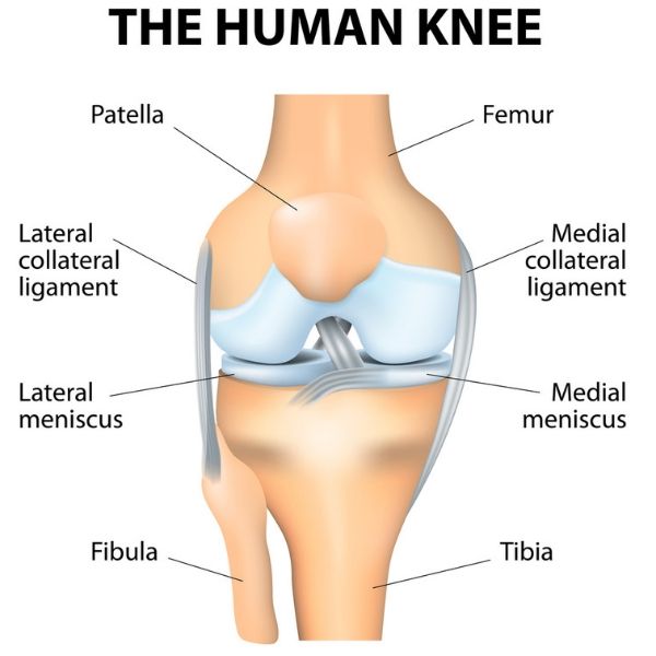
- Three bones meet to form the knee joint, they are: femur (thigh bone), tibia (shin bone), and patella (knee cap.)
- There are two types of cartilage that make up the knee joint:
- The articular cartilage: found on the ends of the femur, tibia and the back of the patella. This shiny, slippery layer acts as a shock absorber and helps the knee bones glide smoothly across each other as the knee is bent or straightened.
- Meniscus: two crescent-shaped discs act as “shock absorbers” and stabilizing structures between the femur and tibia so the bones of the knee can move through their range of motion without rubbing together. Different from articular cartilage, the meniscus is tough and rubbery, containing nerves that help improve stability and balance by assuring the weight is evenly distributed between the femur and the tibia. The meniscus is the only cartilage in the knee that has any circulation. As an adult, this circulation only extends to the outer one third of the meniscus. This is important because it means that tears of the meniscus that extend into the part of the meniscus without a blood supply have no ability to heal. Other meniscus tears in the outer one third can sometimes be repaired and will heal helping them to continue to function and shock absorbers and stabilizers. This also helps to protect the articular cartilage from premature and accelerated breakdown leading to arthritis.
- The knee has two menisci:
- Medial – larger of the two, found on the inner side knee.
- Lateral – found on the outer side of the knee.
- Bones are connected to other bones by ligaments. Ligaments act like strong ropes in the knee, holding the bones together and providing stability. These ligaments are:
- Anterior Cruciate Ligament (ACL): travels from the anterior (front) of the tibia to the posterior (back) of the femur. One of the most important ligaments, the ACL prevents the femur from sliding backward on the tibia, and the tibia from sliding forward on the femur. The ACL the most commonly injured ligament in twisting movements.
- Posterior Cruciate Ligament (PCL): forms an “X” with the ACL ligament, traveling from the posterior (back) of the tibia to the anterior (front) of the femur. The PCL is the largest and strongest ligament in the knee; it prevents the femur from sliding forward on the tibia, or the tibia from sliding backward on the femur.
- The knee has two menisci:
The collateral ligaments stabilize the knee on the inside (medial) and outside (lateral) of the joint and are just outside of the joint (extra-articular) attaching the femur to the tibia. :
- Medial Collateral Ligament (MCL): travels down the inside of the knee and prevents the knee from collapsing inward.
- Lateral Collateral Ligament (LCL): travels down the outside of the knee and prevents the knee from collapsing outward.
- Muscles are connected to bones by tendons. These tough bands of soft tissue provide stability to the knee joint. They are similar to ligaments, but instead of linking bone to bone, they connect muscle to bone. The tendons in the knee that allow extension are:
- Patellar tendon: attaches the patella to the tibia (shin bone.)
- Quadriceps tendon: largest tendon in the knee, connects the muscles in the front of the thigh to the patella.
- While quadriceps and hamstrings are not part of the anatomy of the knee joint, they are important in giving the knee joint strength and flexibility.
- Quadriceps are four muscles in the front of leg that straighten the knee.
- Hamstrings are three muscles in the back of the leg that bend the knee.
- Gluteal muscles: gluteus maximus and gluteus minimus, also called the glutes or the buttocks are important in positioning the knee. The gluteal muscles are also called the hip abductor muscles and are extremely important in the function of the patellofemoral joint. This is the joint behind your patella or knee cap.
- There are approximately 14 bursae in each knee joint. These small, fluid-filled sacs reduce friction between the tissues of the knee and help prevent inflammation.
What are Common Knee Injuries?
The anatomy of the knee is complex, with a lot of moving and working parts. Any of these can be damaged or injured in sporting activities, simple accidents, overuse, or with a degenerative condition. Some of the more common knee injuries treated by Dr. Mazzara are:
- ACL and MCL tears
- Knee Osteoarthritis
- Knee Fractures
- Patella Dislocation
- Patellofemoral pain
- Tendon Ruptures of the Knee
- Ligament Injuries
Many common knee injuries can be treated with a non-surgical approach. If surgery is needed, Dr. Mazzara and his orthopedic team will determine the proper surgical technique to treat the knee injury. Often, minimally invasive surgical techniques can be utilized such as arthroscopic surgery, using a special camera and small surgical instruments.
Work-Related Knee Injuries
To learn more about the anatomy of the knee and the various treatments for knee injuries, please contact the orthopedic offices of Dr. James Mazzara, orthopedic knee specialist in Manchester, South Windsor, Enfield, Glastonbury and surrounding Hartford communities.